
Clinical Problem and Unmet Need
Two of cardiology's most essential diagnostic tools — echocardiography and the 12-lead ECG — share a structural problem: their interpretation quality scales poorly with volume and varies significantly across observers. AI applications in both modalities are designed to address specific, well-documented gaps, though the nature of those gaps differs between them.
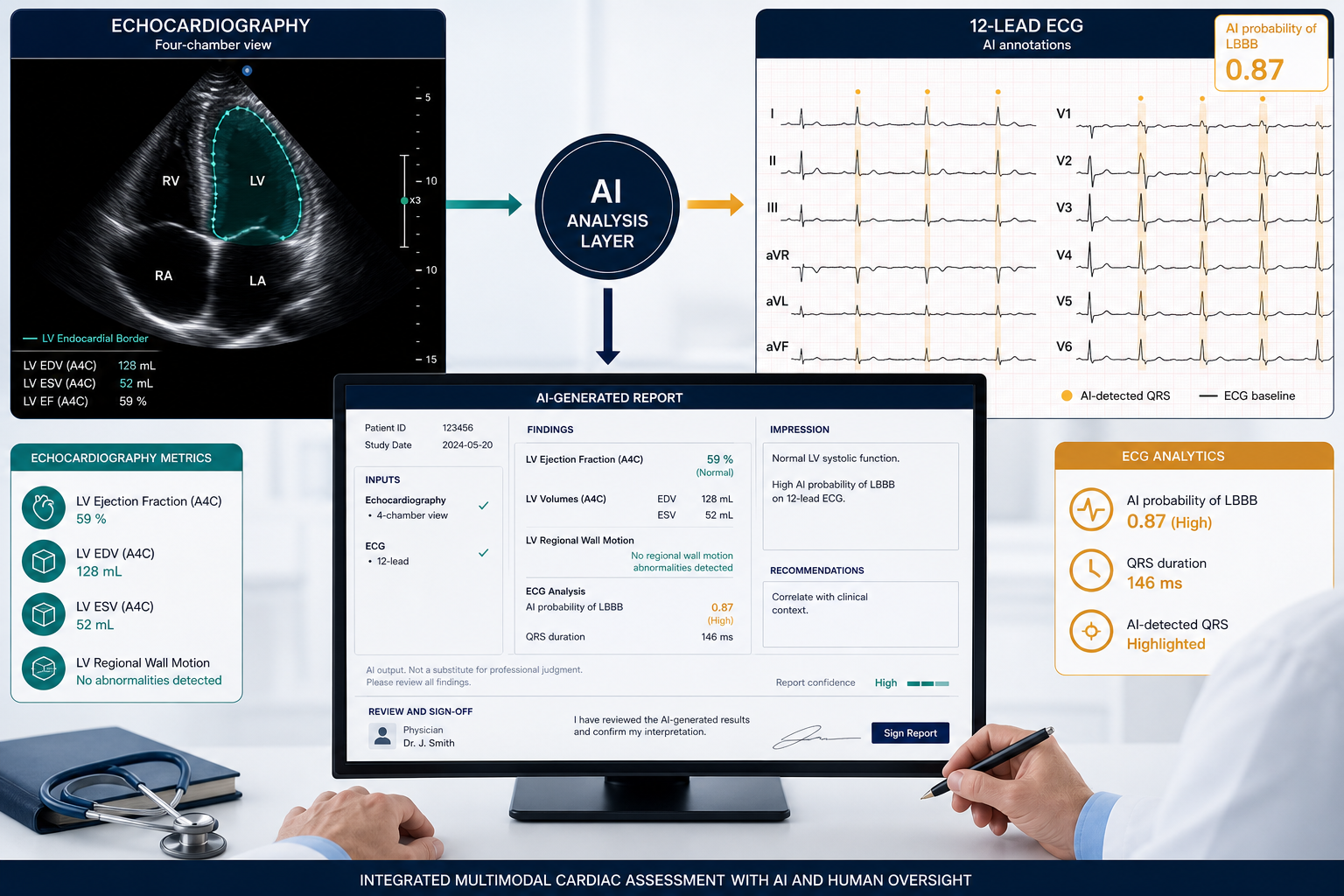
In echocardiography, left ventricular ejection fraction (LVEF) measurement is the most consequential single output of a standard transthoracic study. It drives treatment decisions in heart failure, chemotherapy eligibility, and device implantation. Yet LVEF measurement carries substantial interobserver variability — studies have documented differences of 5 to 10 percentage points between readers, and between a sonographer's initial assessment and the cardiologist's final report. This variability has real clinical consequences: patients near the 35% or 40% threshold for guideline-directed therapy may be classified differently depending on who reads the study and when.
Beyond LVEF, echocardiography faces an access problem. Full transthoracic echocardiography (TTE) requires trained cardiac sonographers, specialized equipment, and cardiologist interpretation — resources that are unevenly distributed. In community hospitals, rural settings, and resource-limited environments, the time from clinical suspicion of cardiac dysfunction to diagnostic imaging can be days to weeks. AI-guided acquisition tools aim to reduce this bottleneck by enabling less-experienced operators to obtain diagnostic-quality images.
ECG interpretation presents a different kind of scaling problem. The 12-lead ECG is ubiquitous — performed hundreds of millions of times annually worldwide — but its diagnostic yield for conditions like reduced ejection fraction or early structural heart disease is limited when read by conventional methods. A normal-appearing ECG does not rule out significant LV dysfunction. AI ECG models, trained on paired ECG-echocardiogram datasets, have demonstrated the ability to detect signals of reduced LVEF, atrial fibrillation, and structural heart disease from the ECG waveform itself — enabling pre-screening at a scale and cost that echocardiography cannot match.
These two modalities are complementary targets for AI, not competing ones. Echo AI addresses variability and quality at the imaging layer; ECG AI addresses scalable pre-screening where imaging is unavailable, delayed, or cost-prohibitive. Understanding both — and the distinct evidence base behind each — is necessary for evaluating the current landscape with appropriate rigor.
The Regulatory Landscape: 510(k) Dominance, Predicate Creep, and the PCCP Pathway
The volume of FDA-cleared cardiology AI devices has grown substantially. According to an analysis of FDA database records by Innolitics, approximately 92 AI/ML-related clearances in cardiology were issued in 2025 alone — up from 62 in 2024 — with 27 manufacturer groups appearing in the cardiology AI clearance landscape for the first time. When counting across both the cardiovascular panel and cardiac-specific imaging AI listed under the radiology panel, the total number of cleared cardiology AI devices reached approximately 203 as of early 2026, according to Cardiovascular Business.
A scoping review published in BMJ Heart in March 2026 analyzed 277 FDA-cleared cardiology AI devices through March 2025 and found that 97.1% were cleared via the 510(k) pathway. This concentration is not inherently problematic — 510(k) clearance requires demonstration of substantial equivalence to a legally marketed predicate device — but the review identified concerning patterns in how predicates are being used.
- 52.2% of cleared cardiology AI devices carried high predicate creep risk — meaning the cleared device's intended use had drifted materially from its predicate's, raising questions about whether the predicate adequately supports the new device's safety and effectiveness claims.
- 50% had a non-AI predicate device — cleared against a conventional (non-AI) device, which may not adequately capture AI-specific failure modes such as model drift or distributional shift.
- 58.8% of clearance summaries underreported the AI technology type — making it difficult for procurers or clinicians to assess the underlying model architecture or its known limitations from public records alone.
Four FDA product codes concentrate most of the echo and ECG AI clearance activity:
| Product Code | Description | Modality |
|---|---|---|
| QYE | Reduced ejection fraction notification software | ECG AI |
| QJU | Guided echocardiographic acquisition | Echo AI |
| QIH | Automated radiological image processing (cardiac) | Echo AI |
| QUO | Echocardiographic quantification software | Echo AI |
The most significant regulatory development of 2025 for this device category is the emergence of the Predetermined Change Control Plan (PCCP) as a governance model. PCCP-authorized clearances allow manufacturers to make pre-specified algorithm updates — within defined bounds — without submitting a new 510(k) for each change. In 2025, 8 of the 92 cardiology AI clearances were PCCP-authorized, the highest single-year count for this category. PCCP-authorized devices include Anumana ECG-AI LEF (K250652, authorized July 2025) and Caption Health Cardiac Guidance (K243065, authorized January 2025). PCCP represents a meaningful shift in how FDA governs iterative AI model improvement, though it also places greater responsibility on manufacturers to define and monitor the bounds of permissible change.
FDA-Cleared AI Tools for Echocardiography
The cleared echocardiography AI landscape spans five distinct functional tasks: LVEF automation, view classification, strain quantification, cardiomyopathy detection, and novice-operator acquisition guidance. The tools below represent the principal cleared platforms as of mid-2026, with regulatory identifiers drawn from FDA records.
| Tool / Company | K-Number(s) | Product Code | Cleared Task(s) | Clearance Date |
|---|---|---|---|---|
| Us2.ai (Us2.ca) | K250151 (plus prior clearances) | SDJ | Automated 2D/Doppler analysis, zero-click structured reporting, cardiac amyloidosis detection, pulmonary hypertension | June 20, 2025 (most recent) |
| InVision Precision LVEF | 510(k) cleared (EchoNet-Dynamic basis) | — | Automated LVEF measurement from TTE | Prior to 2025 |
| InVision Precision Cardiac Amyloid | K243866 | SDJ | Cardiac amyloidosis detection; Breakthrough Device designation; CPT 0932T | May 21, 2025 |
| Caption Health / GE Cardiac Guidance | K243065 | QJU | AI-guided echocardiographic acquisition for novice operators; PCCP-authorized | January 15, 2025 |
| DiA Imaging Analysis — LVivo Software Application | K243862 | — | LV quantification and analysis | 2025 |
| DiA Imaging Analysis — LVivo Seamless | K243331 | — | Automated echo workflow integration | 2025 |
| Ligence Heart | K252105 | QIH | Fully automated TTE analysis and structured reporting | September 26, 2025 |
| Ultromics EchoGo Heart Failure / Core Pro | K222463 (predicate) | QUO | LVEF, strain quantification, heart failure assessment | Prior clearance; active predicate |
Us2.ai represents the broadest deployment footprint in this category, with the company reporting deployment across 500+ hospitals in 28+ countries and over 1 million scans analyzed. Its platform delivers fully automated analysis from DICOM input to structured report without manual measurement steps. The company holds regulatory clearance in 28+ markets and has published validation data in peer-reviewed journals including Nature Communications, Lancet Digital Health, and JACC. These deployment and publication figures are sourced from company-reported information and have not been independently audited for this analysis.
InVision Medical Technology holds two cleared algorithms with distinct clinical profiles. Precision LVEF is the commercial implementation of the EchoNet-Dynamic model developed at Stanford, which was the subject of the only blinded, randomized clinical trial of AI versus sonographer LVEF assessment (discussed in detail in the evidence section below). Precision Cardiac Amyloid carries Breakthrough Device designation and is associated with CPT Category III code 0932T — one of the few echo AI applications with a defined reimbursement pathway as of January 2025.
Caption Health's Cardiac Guidance (now part of GE HealthCare) occupies a distinct niche: it is cleared under product code QJU for guided acquisition, not for automated interpretation. Its intended use is enabling less-experienced operators — nurses, emergency physicians, hospitalists — to obtain diagnostic-quality echocardiographic images using real-time AI feedback. This positions it as an access-expansion tool rather than an interpretation-automation tool, and its PCCP authorization enables GE to update the guidance algorithms within pre-specified parameters.
FDA-Cleared AI Tools for ECG Interpretation
ECG AI clearances in 2025 concentrated heavily on two clinical tasks: detection of reduced left ventricular ejection fraction (LVEF ≤40%) and atrial fibrillation screening. Product code QYE — reduced ejection fraction notification software — received three new clearances in 2025 alone, making it one of the most active product codes in the entire cardiology AI clearance landscape.
| Tool / Company | K-Number | Product Code | Cleared Task | Clearance Date | PCCP |
|---|---|---|---|---|---|
| Anumana ECG-AI LEF | K250652 | QYE | LVEF ≤40% detection from 12-lead ECG | July 28, 2025 | Yes |
| Tempus ECG-Low EF | K250119 | QYE | LVEF ≤40% detection from 12-lead ECG | July 15, 2025 | Not confirmed |
| Bunkerhill ECG-EF | K250649 | QYE | LVEF ≤40% detection from 12-lead ECG | September 19, 2025 | Not confirmed |
| Eko EFAST (Foundation Analysis Software with Transformers) | K251494 | DQD | Heart sound and ECG analysis for murmur and cardiac abnormality detection | August 12, 2025 | Not confirmed |
| Eko Low EF Tool (ELEFT) | K233409 | QYE | LVEF ≤40% detection via stethoscope ECG | March/April 2024 | No |
| Tempus ECG-AF | K233549 | — | Atrial fibrillation detection from 12-lead ECG | Prior to 2025 | Not confirmed |
The Eko Low EF Tool (ELEFT) is notable for its clinical context: it was developed with Mayo Clinic and was the first FDA-cleared AI enabling LVEF ≤40% detection via a stethoscope-based ECG during a routine physical examination, with a 15-second acquisition time. A multi-site prospective validation study of 3,456 patients reported an AUROC of 0.835. An independent validation by Imperial College London published in Lancet Digital Health reported AUROC 0.85, sensitivity 84.8%, and specificity 69.5% in 1,050+ patients, which subsequently prompted NHS deployment across 100+ clinics in London and Wales. This represents one of the clearest examples of a cleared ECG AI tool with independent external validation and documented real-world deployment.
Anumana ECG-AI LEF (K250652) is the most extensively externally validated of the 2025 QYE clearances, with a multisite study published in JACC: Advances in February 2026 (discussed in depth in the evidence section). Its PCCP authorization enables Anumana to update the algorithm within pre-specified parameters, which is particularly relevant for ECG AI given the known sensitivity of these models to ECG device type and sampling rate variation.
Clinical Evidence Quality: What the Prospective Data Actually Shows

The central finding of the BMJ Heart scoping review — conducted by an Imperial College London team and published March 2026, covering 277 cleared cardiology AI devices through March 2025 — is stark: only 3.2% of cleared devices were supported by high-quality multicenter prospective trials. 69.3% were validated by bench studies only. This is not a finding about a few outlier products — it describes the structural condition of the cleared landscape. The 92 new clearances issued in 2025 (after the review's March 2025 cutoff) are not captured in this denominator, so the actual proportion for the full 2025 cohort may differ.
Against this backdrop, three prospective studies stand out as the highest-quality evidence currently available for echocardiography and ECG AI, and each warrants careful interpretation.
The Nature 2023 Blinded RCT: AI vs. Sonographer for LVEF Assessment
The only blinded, randomized non-inferiority trial of AI versus sonographer LVEF assessment was published in Nature in 2023 (He et al., NCT05140642). In 3,495 echocardiographic studies at Cedars-Sinai, the primary endpoint was the proportion of studies where the initial LVEF assessment was subsequently changed by more than 5 percentage points by the reviewing cardiologist. This rate was 16.8% in the AI arm versus 27.2% in the sonographer arm — a difference of −10.4 percentage points, meeting both the non-inferiority and superiority thresholds. Reviewing cardiologists could not reliably distinguish AI-generated from sonographer-generated initial assessments (blinding index 0.088). AI processing also reduced time requirements for both sonographers and cardiologists.
The AI model evaluated in this trial is EchoNet-Dynamic, developed at Stanford and forming the basis of InVision's Precision LVEF product. The trial was conducted at a single academic medical center (Cedars-Sinai) with high-quality TTE studies, and the model was trained on Stanford data — meaning generalizability to community settings, lower-quality image acquisition, or institutions with different equipment profiles is not established by this trial alone. The result is compelling within its scope: AI initial LVEF assessment is non-inferior and superior to sonographer initial assessment on this specific endpoint. It does not establish AI as a replacement for cardiologist interpretation of the full echocardiographic study.
The Anumana JACC Advances 2026 Multisite Validation: ECG AI for Low EF Detection
A retrospective multisite external validation study of the Anumana ECG-AI LEF algorithm was published in JACC: Advances in February 2026. Across 13,960 patients at four geographically diverse US sites — Beth Israel Deaconess Medical Center, Montefiore Medical Center, Monument Health, and the University of Utah — the algorithm achieved:
- AUROC 0.919 for detection of LVEF ≤40%
- Sensitivity 84.5%, Specificity 83.6%
- Negative predictive value (NPV) 98.4% — supporting its use as a rule-out screening tool
- Positive predictive value (PPV) 30.5% at a 7.9% disease prevalence — meaning most positive screens require confirmatory imaging
The NPV of 98.4% is the clinically actionable figure here: a negative Anumana ECG-AI result makes LVEF ≤40% unlikely, suggesting the algorithm could be used to triage which patients require echocardiography for suspected LV dysfunction. The PPV of 30.5% at 7.9% prevalence means that in a general primary care population, roughly two-thirds of positive ECG AI screens would not have confirmed LVEF ≤40% on echo — a clinically important limitation for any screening deployment.
EchoNext (Nature 2025): AI-ECG for Structural Heart Disease Detection
The EchoNext study, published in Nature in July 2025 (Elias et al.), trained a convolutional neural network on 1.245 million ECG-echocardiogram pairs from 8 New York-Presbyterian hospitals. The model's task was detecting a composite of structural heart diseases: LVEF ≤45%, LV hypertrophy, moderate/severe RV dysfunction, pulmonary hypertension, valvular disease, and pericardial effusion.
Internal test AUROC was 85.2%. External validation at Cedars-Sinai, Montreal Heart Institute, and UCSF showed AUROC in the 78–80% range — a 5–7 percentage point drop that reflects real-world generalizability constraints. In a controlled blinded survey, EchoNext achieved accuracy of 77.3% for detecting structural heart disease from ECGs, compared to 64.0% for board-certified cardiologists performing the same task. The DISCOVERY prospective trial (100 patients without prior echocardiography) confirmed a clinically meaningful PPV exceeding 50% for structural heart disease at high-risk EchoNext scores. The model weights and a 100,000-ECG dataset were released publicly.
EchoNext is not currently FDA-cleared as a standalone commercial product. Its significance is methodological and directional: it demonstrates that AI models trained on large ECG-echo paired datasets can detect structural heart disease from ECG waveforms with accuracy exceeding that of cardiologists on this specific task — and that this performance holds across external validation sites, with a quantified but manageable generalizability gap.
| Study | Design | N | Primary Finding | Key Limitation | Funding |
|---|---|---|---|---|---|
| He et al., Nature 2023 (EchoNet/InVision Precision LVEF) | Blinded RCT, single-center | 3,495 echo studies | AI initial LVEF assessment superior to sonographer: 16.8% vs. 27.2% substantial change rate | Single academic center; high-quality TTE only; not generalizable to community settings without further validation | Stanford/Cedars-Sinai (NIH-funded) |
| Anumana ECG-AI LEF, JACC Advances 2026 | Retrospective multisite external validation | 13,960 patients, 4 US sites | AUROC 0.919; NPV 98.4%; PPV 30.5% at 7.9% prevalence | Industry-funded; paced ECGs excluded; PPV low at low prevalence; COI disclosure required | Anumana Inc. (via Mayo Clinic contract) |
| EchoNext, Nature 2025 (Elias et al.) | Internal + external validation + prospective DISCOVERY trial | 1.245M training pairs; external: 3 sites | Internal AUROC 85.2%; external 78–80%; outperformed cardiologists (77.3% vs. 64.0%) on SHD detection | 5–7% AUROC drop on external validation; not FDA-cleared; composite SHD endpoint; cardiologist comparison task was ECG-only (not their typical workflow) | Academic (NYP/Columbia); model weights public |
Known Limitations by Modality
Regulatory clearance establishes that a device is substantially equivalent to a predicate and meets a defined safety and effectiveness threshold for its cleared indication. It does not resolve modality-specific limitations that persist in real-world use. The following limitations are documented in peer-reviewed literature and FDA regulatory records.
ECG AI: Documented Limitations
- Paced ECG exclusion: The Anumana ECG-AI LEF algorithm was trained exclusively on unpaced ECGs. Paced rhythms alter the QRS morphology in ways that invalidate the algorithm's signal extraction, meaning a substantial proportion of patients with cardiac implantable devices — who are often at highest risk for LV dysfunction — cannot be screened with this tool in its current form.
- U-shaped age performance: In the Anumana multisite validation, performance varied significantly by age. The odds ratio for correct detection was 25.8 for patients under 40, 43.4 for patients aged 50–59, and 16.3 for patients aged 80 and older. The model performs least well at the extremes of the age distribution — both young patients (where LV dysfunction is less common and ECG patterns less typical) and very elderly patients (where comorbidities alter ECG morphology).
- Lower specificity in known HF and prior MI patients: ECG AI models trained to detect LV dysfunction show lower specificity in patients who already carry diagnoses of heart failure or prior myocardial infarction — conditions that alter the ECG in ways that may mimic the signal of reduced EF even when current EF is preserved. This limits utility as a monitoring tool in these populations.
- FDA-mandated binary output: Under 21 CFR 870.2380, cleared ECG AI tools for LVEF notification are restricted to binary output — positive or negative for LVEF ≤40%. Continuous probability scores or quantitative EF estimates cannot be presented to clinicians under this regulatory framework, which limits how the output can be integrated into nuanced clinical workflows.
- Low PPV at low disease prevalence: At 7.9% LVEF ≤40% prevalence (representative of a general outpatient population), the Anumana algorithm's PPV was 30.5%. In a lower-prevalence primary care screening context, PPV will be even lower. Most positive screens require confirmatory echocardiography, which must be factored into any deployment's downstream resource implications.
Echo AI: Documented Limitations
- Image quality dependency: AI echocardiography models perform substantially differently on point-of-care ultrasound (POCUS) compared to full diagnostic TTE. Most validated models were trained on full TTE studies from academic centers with high-quality imaging. The 'garbage in, garbage out' principle applies directly: suboptimal image quality from poor acoustic windows, obesity, or POCUS-grade equipment degrades AI output quality in ways that are not always transparent to the operator.
- Training data bias from expert annotation: AI models trained on expert-annotated echocardiographic data inherit the systematic tendencies of those experts. When the same expert or expert group provides both training annotations and serves as the ground-truth comparator for validation, the model has a structural advantage over independent human readers — a methodological limitation that inflates apparent performance in some validation studies.
- Black-box opacity: Most cleared echo AI models do not provide interpretable explanations of their outputs. A cardiologist reviewing an AI-generated LVEF measurement cannot inspect which image features drove the estimate, making it difficult to identify cases where the model may be operating on artifactual signals rather than true cardiac anatomy.
- Limited generalizability to pediatric and congenital populations: Virtually all cleared echo AI tools were trained on adult TTE datasets. Cardiac anatomy in pediatric patients and those with congenital heart disease differs substantially from the adult normal and pathological patterns these models learned. Applying cleared adult echo AI tools in these populations is outside their cleared indications and carries meaningful risk of unreliable output.
Real-World Deployment: Integration, Reimbursement, and Equity
Regulatory clearance and published clinical evidence are necessary conditions for deployment, but they do not resolve the operational constraints that determine whether a tool can actually function in a given clinical environment. The following factors represent the principal real-world deployment barriers as of mid-2026.
EHR and PACS Integration Complexity
Most cleared echo AI tools operate as DICOM post-processing layers — they receive DICOM echo studies from PACS, process them, and return structured measurements or reports. This requires DICOM routing configuration, HL7 or FHIR result delivery back to the EHR, and workflow integration at the ordering, interpretation, and documentation layers. In practice, this means IT project work that is non-trivial in most health systems, particularly those with heterogeneous PACS environments or limited health IT capacity. ECG AI tools face similar integration complexity when deployed in hospital ECG management systems rather than as standalone applications.
CPT Reimbursement Gaps
Reimbursement for AI-specific echocardiography services remains largely unresolved. Most echo AI applications do not have dedicated Category I CPT codes — meaning the AI analysis layer is not separately billable, and health systems must absorb the cost of AI software within existing echocardiography reimbursement. The principal exception is CPT Category III code 0932T, effective January 2025, which applies to AI-assisted cardiac amyloidosis detection — specifically relevant to InVision Precision Cardiac Amyloid. AI-derived fractional flow reserve from CT received Category I CPT codes in 2026, but this applies to coronary CT AI, not echo. The absence of Category I CPT codes for most echo AI applications is a significant barrier to broad commercial deployment in fee-for-service environments.
Model Drift and Post-Market Surveillance
A March 2026 lifecycle governance review published in Frontiers in Digital Health synthesized convergent requirements across US, EU, and UK regulatory frameworks for AI cardiovascular devices. The review identifies continuous post-market surveillance with longitudinal subgroup monitoring as a cross-jurisdictional requirement — but notes that current post-market surveillance practice for cleared cardiology AI devices is immature relative to these expectations. For ECG AI specifically, alert rate stability and calibration across different ECG device types and sampling rates are identified as priority monitoring metrics. For echo AI, image quality-sensitive validation and monitoring are emphasized.
Model drift — the degradation of a model's performance over time as the patient population, clinical workflows, or data acquisition practices shift — is a particular concern for AI tools deployed in dynamic clinical environments. PCCP-authorized tools have pre-specified change control plans, but post-market performance monitoring infrastructure at the health system level remains underdeveloped for most institutions.
Health Equity Concerns
Several equity-relevant performance gaps are documented or plausible for both modalities:
- Race and ethnicity: Most cleared AI tools were trained predominantly on datasets from large academic medical centers, which may not adequately represent the demographic diversity of community hospitals or safety-net institutions. Performance differences across racial and ethnic subgroups are not consistently reported in clearance summaries.
- Age extremes: The U-shaped age performance documented in the Anumana ECG AI validation is a specific, quantified example of a broader concern — AI models may perform differently in pediatric, young adult, and very elderly populations compared to the middle-aged adults who dominate most training datasets.
- Acquisition hardware variation: Echo AI models trained on images from specific ultrasound vendors or probe configurations may perform differently when deployed on equipment from other manufacturers. This is particularly relevant for Caption Health-style guided acquisition tools, where the AI feedback is calibrated to specific image quality parameters.
- Access disparities: AI tools that require high-quality full TTE as input may not reduce access gaps if the limiting factor is sonographer availability, not interpretation quality. ECG AI tools with high false-positive rates in low-prevalence populations may generate downstream echocardiography referrals that strain capacity rather than reducing it.
Evidence-Readiness Summary and Evaluative Framework
The table below maps the principal cleared tools to their evidence tier, known limitations, deployment stage, and key procurement considerations. Evidence tier classification is based on the highest-quality available published evidence, not on FDA clearance status.
| Tool | Modality | Cleared Task | Evidence Tier | Key Limitation | Deployment Stage | Reimbursement |
|---|---|---|---|---|---|---|
| InVision Precision LVEF (EchoNet-Dynamic) | Echo | LVEF automation | Highest: blinded RCT (Nature 2023, n=3,495) | Single-center; high-quality TTE only | Active deployment via PACS/Epic | No dedicated CPT |
| Anumana ECG-AI LEF | ECG | LVEF ≤40% detection | High: retrospective multisite external validation (JACC Advances 2026, n=13,960); industry-funded — COI | Paced ECG exclusion; low PPV at low prevalence; COI | PCCP-authorized; active deployment | No dedicated CPT |
| Eko ELEFT | ECG (stethoscope) | LVEF ≤40% detection | High: prospective multi-site (n=3,456) + independent Lancet DH validation (n=1,050+) | Stethoscope-only ECG; specificity 69.5% | NHS deployment (100+ clinics); US deployment via SENSORA | No dedicated CPT |
| Caption Health / GE Cardiac Guidance | Echo | Guided acquisition (novice operators) | Moderate: prospective usability studies; limited outcomes data | Acquisition quality, not interpretation; POCUS vs. TTE gap | PCCP-authorized; active deployment | No dedicated CPT |
| InVision Precision Cardiac Amyloid | Echo | Cardiac amyloidosis detection | Moderate: Breakthrough Device; limited prospective outcomes data | Specific indication; requires high-quality TTE | Active deployment; Breakthrough Device | CPT 0932T (Category III, Jan 2025) |
| Us2.ai (Us2.ca) | Echo | Automated 2D/Doppler; multi-task | Moderate: peer-reviewed publications; no RCT | Company-reported deployment figures; no independent RCT | 500+ hospitals (company-reported) | No dedicated CPT |
| Ligence Heart | Echo | Full TTE automation and reporting | Limited: cleared Sept 2025; limited prospective evidence published | Very recent clearance; limited post-market data | Early deployment | No dedicated CPT |
| Tempus ECG-Low EF / ECG-AF | ECG | LVEF ≤40%; AF detection | Limited: no independently verified clinical validation data available for this analysis | No public independent validation confirmed | Unknown deployment scale | No dedicated CPT confirmed |
An Evaluative Framework for Procurement
For health systems evaluating these tools, FDA clearance is the floor, not the ceiling, of due diligence. A structured evaluation should address at minimum:
- Evidence quality: Is the validation evidence prospective, multicenter, and externally conducted? Is the study population demographically representative of your patient population? Who funded the study, and are conflicts of interest disclosed?
- Cleared indication scope: What specific task is the tool cleared for, and does that match your intended use? A tool cleared for LVEF ≤40% notification is not cleared for quantitative LVEF reporting or general cardiac screening.
- Known exclusions: Does the tool explicitly exclude paced ECGs, pediatric patients, POCUS-quality images, or other patient subgroups that are common in your clinical environment?
- Integration requirements: What DICOM routing, HL7/FHIR connectivity, and EHR workflow changes are required? What is the IT implementation timeline and resource requirement?
- Post-market surveillance plan: Does the vendor provide performance monitoring data from deployed sites? Is there a defined process for identifying and reporting model drift or unexpected failure modes?
- Reimbursement pathway: Is there a billable CPT code for the AI service in your payer mix, or will the cost be absorbed within existing procedure reimbursement?
- Equity audit: Has the tool been validated in populations similar to your patient demographics? Are subgroup performance data available by race, age, sex, and comorbidity profile?
The PCCP Pathway and Post-Market Surveillance: The Next Governance Phase
The 8 PCCP-authorized clearances in 2025 cardiology AI represent a meaningful governance shift. PCCP enables manufacturers to update algorithms within pre-specified bounds without new 510(k) submissions, which is operationally important for AI tools whose performance can be improved with new training data. However, PCCP also places greater responsibility on manufacturers to define, monitor, and report on the bounds of permissible change — and on health systems to understand what those bounds are when evaluating a PCCP-authorized tool.
The convergence of US, EU, and UK post-market surveillance requirements — as synthesized in the Frontiers 2026 lifecycle governance review — signals that continuous performance monitoring, structured change governance, and longitudinal subgroup tracking will increasingly be expected as baseline practice, not optional add-ons. Health systems that deploy cardiology AI tools without post-market monitoring infrastructure are operating in a governance gap that will become harder to sustain as regulatory expectations mature.
Comments
Join the discussion with an anonymous comment.